The crack echoed across the field, a sickening sound that brought the game to a stop. Once the athlete fell to the ground and held his lower leg. It was obvious that the forces had overwhelmed the very structures that were meant to support and enable movement. It’s in moments like these, or perhaps a simple stumble down the stairs, that we are reminded of the engineering that is underneath our skin.
We often admire the femur, the longest bone in our bodies, or our hand bones, but the tibia and fibula, the hard workers of our lower leg, often go unappreciated until injury strikes. They bear our weight and allow us to move while taking significant loads, so we can walk, run, and jump. Despite their importance, many people don’t understand how the two bones are organized and what they do.
Let’s explore the amazing world of Tibia and Fibula Anatomy with Labeled Diagram that can show us how these bones work together to provide a foundation for everything we do day-to-day. To understand the quality and structure of our lower limb, able to bear heavy loads while being quite fragile, it is important to know our Tibia and Fibula Anatomy with Labeled Diagram.
What are the Tibia and Fibula? Understanding Connection with the Interosseous Membrane?

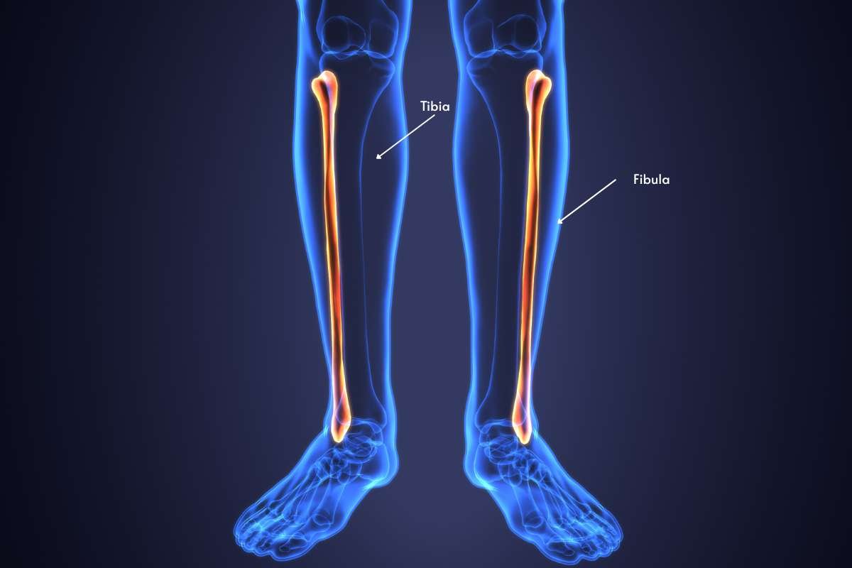
The tibia is the larger and stronger of the two bones. It bears most of the body’s weight when standing or walking. Meanwhile, the Fibula is located on the lateral or outside side of the tibia. It provides support and stability for the ankle joint.
The interosseous membrane acts like a strong, flexible bridge, linking the tibia and fibula together. More than just a connection, this vital sheet of connective tissue plays a crucial role in keeping your lower leg stable, ensuring that weight and forces are distributed evenly between these two essential bones.
Tibia and Fibula Anatomy with Labeled Diagram:
Let’s get more insights about these essential bones in our lower leg.
Tibia Anatomy
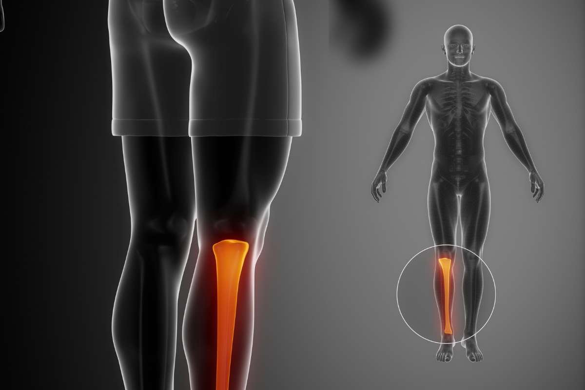
Key features include the prominent frontal crest, the smooth articular surfaces at both ends, the fibular notch at the ankle, and the medial and lateral condyles that shape the knee.
The intercondylar importance steadies the knee, while the medial and lateral malleolus strengthen the ankle. Finally, the tibial tuberosity provides an essential anchor for muscles and ligaments.
Here’s a breakdown of the different parts of the tibia:
- Anterior crest: Sharp ridge on the anterior (front) surface of the tibia
- Articular surface of the tibia: A smooth surface on the proximal end that articulates with the femur
- Fibular notch: Concave surface on the lateral side of the tibia that is part of the ankle joint
- Medial and lateral condyles: Form the inner and outer parts of the knee joint
- Articular surface of medial and lateral condyles: Smooth, articulating surfaces that promote movement in the knee joint
- Intercondylar eminence: A ridge between the two condyles, which helps to stabilise the knee joint
- Medial and lateral malleolus: Form the inner and outer parts of the ankle joint, providing stability
- Tibial tuberosity: A bony protrusion on the anterior surface that provides a point for muscles and ligaments to attach to.
Fibula Anatomy
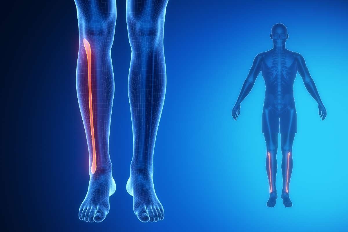
The shaft provides numerous attachment points for muscles that move your ankle and foot. Down at the ankle, the distal end forms the noticeable lateral malleolus, a key structure for stabilizing the ankle joint and anchoring important ligaments.
Here are some key features of each part:
- Proximal end: The proximal or upper end of the fibula is commonly called the head. The head is circular and makes contact with the lateral condyle of the tibia to function in the knee joint. This joint permits rotation and gliding of the knee.
- Shaft: The fibula has a long and thin shaft that is slightly curved. The shaft serves as the attachment site for muscles that function in movements of the ankle and foot.
- Distal end: The lateral malleolus is the distal or lower end of the fibula. It forms the bony protrusion on one side. The opening of the ankle that prevents excessive eversion.
After reading this, you can understand well about Tibia and Fibula Anatomy with Labeled Diagram.
What’s Orthopaedics’ Take on Tibia and Fibula Fracture?
After understanding Tibia and Fibula Anatomy with labeled diagram and considering their vital role in support and movement. Protecting your tibia and fibula is important, as injuries or fractures can severely impact mobility. Let’s learn more about how to safeguard these essential bones.
Fractured Tibia and Fibula
A tibia-fibula fracture occurs when a sudden fall or forceful blow overwhelms the lower leg bones, causing them to break. This is a serious injury demanding immediate medical attention, but with prompt and appropriate treatment, a full recovery is often possible.
Types of Tibia and Fibula Fractures
1. Cozen’s fractures
They are most prevalent under the age of six. Cozen’s fractures occur at the top of the tibia when a great deal of force is applied to the side of the knee, causing a bending moment or force. An example would be when a young child takes themselves off a trampoline foot-first and lands off or sideways and gets their leg trapped underneath their body while going down the slide or sledding.
2. Toddler fractures
They occur in young children, typically under the age of four. They are often caused while the young child is ambulating and falling or stumbling by twisting their leg– this is how a toddler fracture occurs. They happen in the middle of the tibia and can be hard to see on an X-ray.
3. Tibial tubercle fractures
It occurs in adolescence. The tibial tubercle is a bony bump on the upper portion of the shin where the quadriceps muscle attaches to the bone via the patellar tendon. Tibial tubercle fractures are a break or crack in this position. This type of injury occurs when the child’s tibial tubercle is going through the growth process, and the bone in the area is soft.
Also Read: Understanding the Anterior Tibialis Muscle: Anatomy, Function, and Common Injuries
Symptoms & Causes
A tibia-fibula fracture’s symptoms are:
- Pain or swelling in the lower leg
- Inability to stand or walk, this is less likely if only the fibula is broken
- Limited range of motion in the knee or ankle area
- Bruising or discoloration of the skin around the break
Fractures of the tibia and fibula are often from a fall or a hard blow to the leg that applies excessive force to the bone. Common causes are:
- A sudden twist of the leg is stiff or planted in place, which is common in football, hockey, and basketball
- Falls while ice skating, skiing, or snowboarding, when the foot is secured in a boot, the fracture often occurs above the boot
- Falls on a trampoline or playground structure
Source: https://www.childrenshospital.org/conditions/broken-tibia-fibula-shinbonecalf-bone#diagnosis–treatments
Clinical Trial: Fibular notch morphometry and its clinical importance on dry bones
In a thorough morphometric examination of 76 dry tibial bones, there emerged a review of the fibular notch anatomy – a critical anatomical feature for fibular and tibial articulation related to ankle stability. Documented assessments of fibular notch depth, fibular notch width, fibular facet angles, and depth are important measurements that directly influence surgical treatments, fracture management, and radiological assessments of the distal tibiofibular region. When represented with labeled diagrams of the tibia and fibula, understanding these features, especially in regards to ankle injury diagnoses and eventual ankle reconstruction plans, is critical for clinicians.
Read the full study here: https://journals.plos.org/plosone/article?id=10.1371/journal.pone.0307387
Conclusion
The sudden possibilities of a sports injury to the complex patterns of everyday motion, the tibia and fibula are essential to the function and endurance of our lower limb. Knowing the details of Tibia and Fibula Anatomy with Labeled Diagram will allow us to understand their impressive design and its fragility. Whether it is identifying the signs of a fracture or simply contemplating the marvel of the body’s engineering, the recognition of Tibia and Fibula Anatomy with Labeled Diagram is your first step to better care and respect for these largely forgotten supports hiding beneath our skin.
FAQ:
1. How long does a broken tibia and fibula take to heal?
A broken tibia and fibula typically takes 3 to 6 months to heal, but this can vary based on the severity of the fracture and the treatment method used. Some severe fractures may take longer, even up to a year or more, for a full return to sports and high-impact activities.
2. Can a broken tibia and fibula heal without surgery?
In some cases, it’s possible to treat a tibia fracture without surgery. A nonsurgical approach may be considered if the break is minor and does not extend into a joint. It may also be an appropriate choice for a patient who is less active or who has health problems that could complicate surgery or recovery.
3. What is the best treatment for a tibia and fibula fracture?
Immobilization: If the break is mild and your bones didn’t move far out of place (if it’s non-displaced), you might only need a splint or cast. …
Closed reduction: More severe breaks require a closed reduction to set (realign) your bones.